
The agenda ![]() is now available
is now available
Projectors for talks use a 16:9 screen format
Poster presentations ![]() will have to be in A0 vertical format!
will have to be in A0 vertical format!
The List of Participants is now available
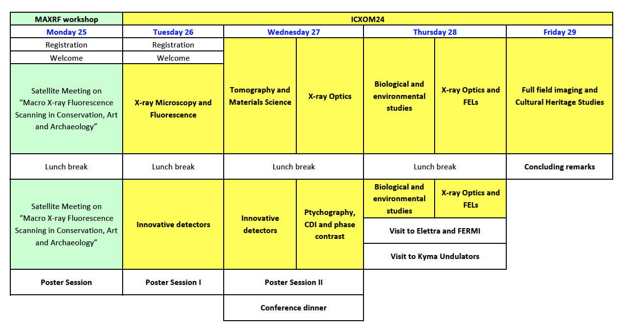
Monday, 25 September 2017
MA-XRF workshop
Tuesday, 26 September 2017
ICXOM24 conference
08:15 08:45 |
Registration |
Welcome
Chair: Alessandra Gianoncelli (Elettra Sincrotrone Trieste)
Room: Oceania ABC
| 08:45 |
Welcome |
| 09:00 09:10 |
Publishing with Nature Communications |
X-ray Microscopy
Chair: Maya Kiskinova (Elettra Sincrotrone Trieste)
Room: Oceania ABC
| 09:10 |
I08-SXM: The Scanning X-ray Microscopy Facility at the Diamond Light Source |
| 09:50 |
A scanning soft x-ray microscope in absorption and fluorescence modes at the Pohang Light Source (PLS) |
| 10:10 |
Nanoscale Multimodal Imaging Capability of the Hard X-ray Nanoprobe at the NSLS-II |
| 10:30 10:50 |
X-ray phase-contrast imaging and metrology using periodic and random wavefront modulators |
| 10:50 11:20 |
Conference picture & Coffee break |
X-ray Microscopy and X-ray Fluorescence
Chair: Maya Kiskinova (Elettra Sincrotrone Trieste)
| 11:20 |
X-ray Fluorescence Imaging and Spectroscopy with High Spatial Resolution at NSLS-II |
| 12:00 |
Analytical requirements for quantitative X-ray fluorescence nano-imaging of metal traces in solid samples |
| 12:20 |
Focusing and Diffraction Properties of Micro-Channel Plates for Transmitted X-ray Radiation |
| 12:40 13:00 |
Nanotomography and X-Ray Fluorescence Microscopy for quantitative Iron concentration map in inflamed cells |
| 13:00 14:50 |
Lunch |
Detectors
Chair: Ralf Menk (Elettra Sincrotrone Trieste)
Room: Oceania ABC
| 14:50 |
Multi-element Germanium Detectors for Synchrotron Applications |
| 15:30 |
ARDESIA: 4-Channels Fast SDD X-ray Spectrometer for Synchrotron Applications |
| 15:50 16:10 |
Seven-Element SDD with Optimized Packing Factor to Reduce Scattering Background |
| 16:10 16:40 |
Coffee break |
Detectors and Instrumentation
Chair: Ralf Menk (Elettra Sincrotrone Trieste)
Room: Oceania ABC
| 16:40 |
X-ray microanalytical activities at the IAEA Nuclear Science and Instrumentation Laboratory |
| 17:00 17:20 |
Fast switching between 2D and direct, 3D XRF imaging using Collimating Channel Arrays and the Maia detector |
| 17:10 19:00 |
Drinks and Poster Session |
Wednesday, 27 September 2017
ICXOM24 conference
Tomography
Chair: Gerd Schneider (Helmholtz-Zentrum Berlin, Germany)
Room: Oceania ABC
| 08:30 |
Time resolved X-ray diffraction computed tomography for studying real systems under operando conditions |
| 09:10 |
Evaluation of the XTM setup at the P05 Imaging Beamline at PETRA III using a sample of Nanoporous gold |
| 09:30 |
Extreme Imaging with the synchrotron pink beam |
| 09:50 |
Inspecting sub-grain level deformation in poly-crystals |
| 10:10 10:30 |
P03 nanofocus end-station for material science |
X-ray Optics
Chair: François Polack (Soleil, Paris, France)
Room: Vulcania I
| 08:30 |
Diffractive X-ray Optics for Synchrotrons and Free Electron Lasers |
| 09:10 |
Fresnel zone plates for nanoARPES |
| 09:30 |
Imaging with Nanometer Resolution from 8 to 100 keV using Multilayer Zone Plates (MZP) |
| 09:50 |
Fabrication and characterization of monolithic multilayer Laue lens nanofocusing optics for hard x-ray microscopy |
| 10:10 10:30 |
Development of X-ray refractive optics for new diffraction limited X-ray sources |
| 10:30 11:00 |
Coffee break |
Tomography and Materials Science
Chair: Lucia Mancini (Elettra Sincrotrone Trieste)
Room: Oceania ABC
| 11:00 |
Visualizing batteries discharge products at Mistral |
| 11:20 |
3-D structure of a gasoline spray by polycapillary X-ray micro-tomography |
| 11:40 |
Three-dimensional characterization of magnetic multilayer thin films using resonant soft X-ray scattering |
| 12:00 12:20 |
Hard X-ray in-situ full-field microscopy for material science |
Data Analysis for XRF and CT
Chair: Chris Ryan (CSIRO, Australia)
Room: Vulcania I
| 11:00 |
Fully-fractionated microprobe analysis |
| 11:20 |
Matrix Factorization for the near real time analysis of XRF imaging data |
| 11:40 |
Analyzing 4D Tomographic X-Ray Spectromicroscopy Data with Mantis |
| 12:00 12:20 |
Monochromatic beam based quantitative dynamic micro computed tomography |
| 12:20 14:00 |
Lunch |
Detectors and Instrumentation
Chair: Peter Siddons (BNL, USA)
Room: Oceania ABC
| 14:00 |
Advances in hybrid pixel detectors for photon science |
| 14:40 |
EIGER and PILATUS3 CdTe detector systems for advanced X-ray studies |
| 15:00 |
Maia Mapper: High definition XRF imaging in the lab |
| 15:20 |
High Speed, Simultaneous XRD-XRF Mapping |
| 15:40 |
The Munich Compact Light Source: Performance upgrades and biomedical research |
| 16:00 |
Micro-X-Ray fluorescence spectrometer with X-ray single bounce gold capillary optics for light element analysis |
| 16:20 16:40 |
Volume Elemental Mapping in Confocal Geometry for Spectroscopy Studies of Biological Samples |
Ptychography and Phase Contrast
Chair: Georgios Kourousias (Elettra Sincrotrone Trieste)
Room: Vulcania I
| 14:00 |
Is the future bright for simultaneous X-ray ptychography and fluorescence microscopy? |
| 14:40 |
Higher dimensional ptychography at the ALS |
| 15:00 |
Simultaneous XRF and Ptychographic imaging of FCC Particles |
| 15:20 |
Understanding the structure and aging of Zwischgold |
| 15:40 |
Ultra high-speed X-ray phase-contrast imaging using single-pulse synchrotron radiation |
| 16:00 16:40 |
(Resonant) Hard X-ray Ptychography for High-Sensitivity Imaging with Chemical Contrast |
| 17:10 19:00 |
Coffee break and Poster Session |
Thursday, 28 September 2017
ICXOM24 conference
Life Sciences
Chair: Gerald Falkenberg (DESY, Germany)
Room: Oceania ABC
| 08:30 |
Elemental imaging at micro and nanoscale in toxicology research: from occupational diseases to reproductive medicines |
| 09:10 |
X-ray fluorescence imaging as a tool for recognition of different types of the ovarian cancer tissues |
| 09:30 |
Characterization of cortical bone demineralization by X-ray-based techniques: A micro and nano scale study |
| 09:50 |
X-ray based micro- and nanoimaging of nanoparticles in exposed biota |
| 10:10 10:30 |
Investigating differences in Nanoscale distribution of trace metals in biominerals |
X-ray Optics and FEL
Chair: Marco Zangrando (Elettra Sincrotrone Trieste)
Room: Vulcania I
| 08:30 |
X-ray optics, state of the art and current issues |
| 09:10 |
Stitching capabilities at the LCLS Metrology Laboratory |
| 09:30 |
Diagnostic of Forward Bragg Diffraction Hard X-ray beams for Self-Seeding applications SwissFEL |
| 09:50 |
Hard X-ray interferometers fabricated by Si planar technologies |
| 10:10 10:30 |
Wavefront sensing using ptychography at FELs |
| 10:30 11:00 |
Coffee break |
Life Sciences and Environment
Chair: Lorella Pascolo (University of Trieste, Trieste, Italy)
Room: Oceania ABC
| 11:00 |
Imaging of chemical composition in plant tissues: revealing physiological mechanisms to improve food quality and safety |
| 11:40 |
Confocal X-Ray Fluorescence Microscopy at the Advanced Photon Source Sector 20 |
| 12:00 |
A first look at the quantification capabilities of the prototype Mars 2020 Planetary Instrument for X-ray Lithochemistry |
| 12:20 12:40 |
QESA - Quarantine Extraterrestrial Sample Analyses: methodology and setup |
X-ray Optics and FEL
Chair: Christian David (PSI, Switzerland)
Room: Vulcania I
| 11:00 |
Diffractive/Reflection optical elements. From Bragg reflection to total external reflection |
| 11:20 |
Wavefront preserving mirrors at LCLS |
| 11:40 |
Development of kinoform lenses made of quartz for microbeam X-ray diffraction |
| 12:00 |
Coated Hollow Capillaries as X-Ray Optics |
| 12:20 12:40 |
Miniaturized X-ray zoom lens |
| 12:40 |
Lunch |
Optics system
Chair: Ralf Menk (Elettra Sincrotrone Trieste)
Room: Vulcania I
| 14:15 |
Optimized High Energy Performance of Polycapillary Optics for µXRF Analysis |
| 14:30 |
Polycapillary Optic for Liquid-Metal-Jet X-Ray Tubes |
| 14:45 |
Optimized High Energy Performance of Polycapillary Optics for µXRF Analysis |
| 15:00 |
Visit to Elettra and Kyma |
Friday, 29 September 2017
ICXOM24 conference
Full-field and tomography
Chair: Juergen Thieme (NSLS-II, USA)
Room: Oceania ABC
| 08:30 |
Nanoscale spectroscopy and tomography with the HZB full-field transmission X-ray microscope |
| 09:10 |
Beam tracking phase tomography with laboratory sources |
| 09:30 |
Comparison of CCD and sCMOS Detector for Sub-micron Resolution in Lab-based X-ray Computed Tomography |
| 09:50 |
X-rays, microscopy, and rock and roll |
| 10:10 10:30 |
The Full-Field XRF Imaging System For Investigation Of Paintings |
| 10:30 11:00 |
Coffee break |
Cultural Heritage
Chair: Franco Zanini (Elettra Sincrotrone Trieste)
Room: Oceania ABC
| 11:00 |
Discrimination of organic compounds in ancient and historical materials with in situ synchrotron-based X -Ray Raman scattering |
| 11:20 |
Monte Carlo simulation and EDXRF analysis of the pigments used in “Natura Morta” (1946) by Mario Mafai |
| 11:40 |
Synchrotron XRF/XAS Applications on Heritage glasses in Thailand |
| 12:00 |
The Bernstorf Gold u2013 A Challenge to the Flexibility of SRXRF |
| 12:20 12:40 |
In-air PIXE, MeV-SIMS, and DAPNe-MS: a multidimensional analytical approach for unlocking chemical information in scalp hair |
Room: Oceania ABC
| 12:40 13:00 |
Concluding remarks |
Confirmed Invited Speakers
The following scientists have already agreed to give invited talks:
Anna Bergamaschi (PSI, Switzerland) - Advances in hybrid pixel detectors for photon science ![]()
Christian David (PSI, Switzerland) - Diffractive X-ray Optics for Synchrotrons and Free Electron Lasers
Burkhard Kaulich (Diamond Light Source, UK) - I08-SXM: The Scanning X-ray Microscopy Facility at the Diamond Light Source ![]()
Marco di Michiel (ESRF, France) - Time resolved X-ray diffraction computed tomography for studying real systems under operando conditions ![]()
Lorella Pascolo (University of Trieste, Trieste, Italy) - Elemental imaging at micro and nanoscale in toxicology research: from occupational diseases to reproductive medicines ![]() '
'
François Polack (Soleil, Paris, France) - X-ray optics, state of the art and current issues ![]()
Juliane Reinhardt (DESY, Germany) - (Resonant) Hard X-ray Ptychography for High-Sensitivity Imaging with Chemical Contrast ![]()
Gerd Schneider (Helmholtz-Zentrum Berlin, Germany) - Nanoscale spectroscopy and tomography with the HZB full-field transmission X-ray microscope ![]()
Pete Siddons (BNL, USA) - Multi-element Germanium Detectors for Synchrotron Applications ![]()
Juergen Thieme (NSLS-II, USA) - X-ray Fluorescence Imaging and Spectroscopy with High Spatial Resolution at NSLS-II ![]()
Michael Jones (Queensland University of Technology, Brisbane, Australia) - Is the future bright for simultaneous X-ray ptychography and fluorescence microscopy? ![]()
Katarina Vogel Mikuš (University of Ljubljana and Joseph Stefan Institute, Slovenia) - Imaging of chemical composition in plant tissues: revealing physiological mechanisms to improve food quality and safety ![]()
....